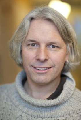
Richard Neutze took his PhD in Physics in 1995 from the University of Canterbury (New Zealand). He was introduced to molecular biophysics at Oxford University (England); completed a postdoc at Tübingen University (Germany); and then moved to Uppsala University (Sweden). In 1998 Neutze became Assistant Professor at Uppsala University and he moved his group to Chalmers University of Technology in 2000. In 2006 Neutze was appointed Professor of Biochemistry at the University of Gothenburg. He has worked on the structural biology of aquaporins; bacterial rhodopsins; photosynthetic reaction centres; and time-resolved diffraction and time-resolved wide angle X-ray scattering studies of membrane proteins, using both synchrotron radiation and X-ray free electron lasers. https://scholar.google.se/citations?user=TLpr7bIAAAAJ&hl=en
Venue: LINXS Workshop room, Ideon Delta 5, Scheelevägen 19, 5th floor (map)
Time: 13.30 - 14.30 (Coffee and cake is served after the seminar)
Title: Time-resolved serial femtosecond crystallography studies at X-ray free electron lasers reveal structural changes in bacteriorhodopsin
Abstract:
Eriko Nango, Tomoyuki Tanaka, Takanori Nakane, So Iwata, Kyoto University & Riken SPring-8 Center (SACLA), Japan.
Przemyslaw Nogly, Tobias Weinert, Jörg Standfuss, Gebhard Schertler, Paul Scherer Institute, Switzerland.
Antoine Royant, ESRF, France.
Cecilia Wickstrand, Richard Neutze, University of Gothenburg, Sweden.
X-ray free electron lasers (XFELs) provide a billion-fold jump in the peak X-ray brilliance when compared with synchrotron radiation. One area where XFEL radiation is having an impact is time-resolved structural studies of protein conformational changes. I will describe how we used time resolved serial femtosecond crystallography at an XFEL to probe light-driven structural changes in bacteriorhodopsin. Bacteriorhodopsin is a light-driven proton pump which has long been used as a model system in biophysics. The mechanism by which light-driven isomerization of a retinal chromophore is coupled to the transport of protons “up-hill” against a transmembrane proton concentration gradient involves protein structural changes. Collaborative studies performed at SACLA (an XFEL in Japan) and the LCLS (an XFEL in Stanford, USA) have probed structural changes in bacteriorhodopsin microcrystals on a time-scale from femtoseconds to milliseconds. Structural results from these studies enabled the photoisomerization of retinal to be visualized (Nogly et al. 2018) and a complete picture of structural changes during proton pumping by bacteriorhodopsin to be recovered (Nango et al., 2016).
Nango, E., et al. A three-dimensional movie of structural changes in bacteriorhodopsin. Science 354, 1552-1557 (2016).
Nogly, P. et al. Retinal isomerization in bacteriorhodopsin captured by a femtosecond x-ray laser Science, eaat0094 (2018)


