ABOUT
The overarching goal of the Advanced Imaging and Data Analysis (AIDA) theme is the formation of a sustainable, long-term funded Nordic research programme and the establishment of a physical hub and competence platform that can continue developing imaging techniques for the future. In this way, we will establish a network for fostering advanced imaging experts through building excellence and innovation in both research and education.
The Theme builds upon the unique expertise gained in two previous LINXS themes (Imaging and Northern Lights on Food (NLF)) and aims to promote the continued advancement and refinement of both sample preparation and instrumentation, fostering development of scientific and innovation environments to enable cutting-edge deep technology and learning across science areas. This integrated approach of method and science areas will create a long-term benefit to the overall user community both with a continuous stream of new students and new groups, as well as building on the people that have already taken their first steps on the imaging path.
HAPPENING IN THEME
CORE GROUP
WORKING GROUPS FOR AIDA
AIDA working group 1
Spectral Nano-imaging
In this working group, the chemical speciation takes centre stage in combination with ultimate resolution methods in chemical imaging. This means, for instance, identifying elements with X-ray Fluorescence, but also disentangling different chemical species of the same element with Scanning Transmission X- ray Microscopy and complementary IR-microscopies. Mostly conducted in 2D, the 3rd dimension is formed by the spectral component. The big advantage is that these techniques require no labelling and instead purely work with the composition or chemistry inherent to the sample, which is therefore relevant for a wide variety of scientific fields. Over the past five years, nano-imaging techniques have come to fruition at MAX IV, with the addition of new operational beamlines and end-stations, and now the time is right for e.g., the development of more streamlined data analysis approaches and assistance with sample preparation for experiments. A separate aim is to reach out to experts in laboratory-based, high-resolution imaging techniques, including optical (fluorescence) methods and spatial proteomics as well as electron-based methods such as TEM and STM while bridging to working group 2. This integration expands the scope of synchrotron experiments, thus providing a comprehensive nano-imaging toolkit.
AIDA WORKING GROUP 2
Micro-tomographic Imaging
Phase-contrast micro-computed tomography is an imaging modality that has evolved rapidly in the past few decades. The method exploits the wave-like behaviour of x-rays and neutrons, providing images with superior contrast in comparison to standard attenuation-based imaging. Due to the coherent properties of synchrotron radiation, phase-contrast imaging has been implemented at all synchrotron imaging beamlines, but even with neutron imaging it is possible to obtain phase-contrast images by use of optical gratings to produce beam interference. In these grating interferometers, it is even possible to extract so-called dark-field images based on the scattering in the sample. Such grating interferometers are even implemented at laboratory x-ray micro-tomography setups, along with propagation-based phase-contrast imaging.
The method experts in WG2 of the AIDA theme work with micro-tomographic imaging at synchrotrons, at neutron facilities, at free-electron laser facilities, and at laboratory-based x-ray sources both in academia and industry. Networking activities internally in the WG2 will provide cross fertilization for method optimization at different instruments and provide a strong basis of competence for the Advanced Study Groups.
AIDA WORKING GROUP 3
Advanced image processing
AIDA advanced study GROUP:
tissue and in vivo imaging
AIDA advanced study GROUP:
food and biotech
AIDA advanced study GROUP:
paleontology and geotech
This working group is a fundamental layer between the acqiusition of raw data and scientific interpretation, dealing with specific data-oriented goals while facing a cross-disciplinary challenge. Besides focused activities inside the working group to advance the use of e.g. machine learning, VR and AR for imaging methods, cross-working group initiatives, such as hackathons and visualization-themed workshops are also foreseen, organized around the central themes provided by the Advanced Study Groups.
The primary emphasis in this working group will be on innovative approaches for extracting multiscale information from X-ray/neutron imaging data, visualizing results in an comprehensive way and delivering this whole process as a user-friendly environment. Additionally, we aim to foster collaboration among Imaging method users to share their expertise to educate and mentor the next generation of scientists.
Tissues are inherently three dimensional, yet the traditional way of analyzing different types of biological and pathological processes in tissue is to section it and then apply various methods like histology, immunohistochemistry and in situ hybridization on the thin 2D slices. However, many organs contain structures which may be difficult to understand from a 2D slice. This is especially the case when the tissue organization has been disrupted by disease processes. During recent years, life science researchers from different fields have started to explore the use of synchrotron-based X-ray imaging methods such as phase-contrast micro-CT, X-ray fluorescence mapping and scanning SAXS/WAXS for tissue characterization. Since the imaging can be performed in a non-invasive way, valuable tissue from biobanks can be scanned and analyzed. Currently there is a great need for knowledge exchange between researchers from different fields to make sure that novel techniques are shared and used in the most efficient way.
Biomass and derived food materials are complex assemblies of biopolymers and small molecules, in terms of their molecular composition and multiscale organization from the molecular to the macroscopic level. Moreover, all processing steps in industrial biotechnology and food (bio)technology influence this multiscale structure, and they often require time resolved analyses to follow their development. Considering the food sector, all steps in the preparation of the food products, from the raw materials, extraction of suitable ingredients, processing and storage, affect the sensory properties of the food we ingest as consumers, the bioavailability of the nutrients through the gastrointestinal tract, and the derived health benefits. Therefore, the integration of imaging techniques coupled with X-ray and neutron techniques is required to understand the composition and structural properties of food materials in all steps of their cycle. Efforts should be made to develop new tools for image processing and analysis of the vast amount of data generated by such experiments with the help of deep machine learning and artificial intelligence tools.
Geological, fossil and archaeological samples form a large component of our cultural heritage and, in that sense, they can be far too rare and precious to be sectioned or sampled. Three-dimensional imaging has been developed to overcome this limitation and allow for microstructural and chemical analysis of this crucial material. However, this is not without challenges. These samples are often dense and comprise embedded metallic oxides that strongly react with X-ray and create artifacts. We have formed a community of synchrotron scientists and synchrotron users (with key members in the Nordic countries) to develop and apply synchrotron X-ray imaging on these samples. We propose to introduce the methods of X-ray (micro-)tomography, diffraction tomography and fluorescence to the communities of Geotech and Cultural Heritage to increase the visibility of these techniques to the study of our Nordic history.









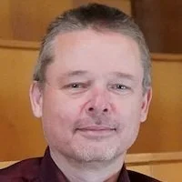


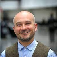


Advanced Imaging and Data Analysis: Core Group Leader, LINXS Fellow
Core responsibility in Theme: Education, Competence, and Innovation
Associate Professor, Department of Medical Radiation Physics, Lund University, Sweden.
Martin is Associate Professor at Medical Radiation Physics at Lund University. He has a scientific background in X-ray physics and specialized in X-ray imaging method development. Martin has a strong track record of grating-based X-ray interferometry for X-ray phase-contrast and X-ray dark-field imaging for imaging of organic materials at synchrotron radiation facilities, at conventional X-ray sources and at neutrons facilities. He has been an active member of the LINXS community since the start of the Imaging theme and is playing an active role in the development of a new beamline at MAX IV dedicated to tomography and imaging of organic materials. He has more than 10 years of experience in undergraduate teaching of ionising radiation and was co-founder and leader of the graduate research school “Imaging of 3D structures” organised across the faculties of science, engineering, and medicine at Lund University 2014-2019. In addition to user experience at multiple European synchrotrons, Martin is engaged in the proposal review panels at PETRA III and MAX IV. He has received research funding from the Swedish Foundation for Strategic Research, from the Swedish Research council, and is currently PI on an ERC consolidator grant developing X-ray imaging instrumentation for bio-medical research.