French master students spend summer in a unique internship using X-ray microtomography
For four weeks in June, two French master students in Microbiology and Physiochemistry applied to food and wine processes, (International Master’s programme MP2) from the “Université Bourgogne Franche Comté” (UBFC, Dijon, France), spent a unique internship at the Division of Solid Mechanics, Department of Construction Science and at the Department of Food Technology at Lund University (LU). The students could bring back learnings of how to use X-ray microtomography and image processing to France and will thus broaden the network of future users of these technologies.
During their stay they were supervised by researcher Emanuel Larsson, member of two themes within LINXS – the Northern Light on Foods (WP3) and the Integrative Pharmacology and Drug Discovery (WP3):
– This unique opportunity for the students comes from a personal and long collaboration with Professor Camille Loupiac at Institut Agro Dijon, France who has also been a strong personal influence on how I came to start with neutron tomography, says Emanuel Larsson.
Today he is a method expert in X-ray and Neutron imaging at the Division of Solid Mechanics and an application expert in tomographic imaging and image analysis at LUNARC - Center for Scientific and Technical Computing.
During the last couple of years, Professor Loupiac has invited Emanuel each summer to hold lectures at Institut Agro Dijon and UBFC together with demo sessions of his Kitchen-Based Light Tomography (KBLT) scanner. There, he has presented real life examples of X-ray and neutron tomography of bread and food science samples. Earlier this year, she asked if this exchange could also be extended to an internship for two Master students, Sahak Yeghiazaryan and Theó Pointurier.
Analysing bread samples with high nutritional contents
The aim of the internship was for the two students to learn how to use various technologies and to analyse data from their own samples. Prior to the internship they set up a replica of the KBLT scanner, but now they also got the opportunity to use real X-rays instead of visual light. Normal light is sufficient to study the inner parts of samples that are transparent to the human eye, whereas X-rays can often provide insights of the inside of non-transparent material.
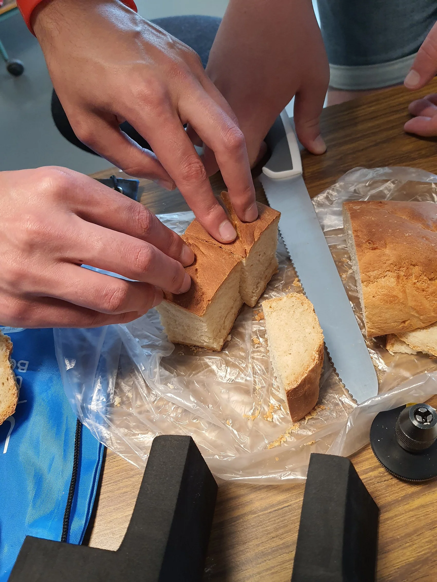
– Our project this year is to formulate a bread with spirulina algae. In France we focused on the formulation and texture of the bread. Here in Lund, we have been baking bread and then scanned it with the X-ray microtomography scanner to create 3D volumes, says Sahak Yeghiazaryan.
Examining the texture is important as this is one of the most important factors when consumers choose which bread to buy. The choice of studying bread made with spirulina algae is that it also has a lot of nutritional benefits with a high content of proteins and contains several minerals and vitamins, which are other important market aspects.
– We are interested in the texture, especially the porosity, which we can investigate with X-ray microtomography in a non-destructive manner, without having to cut the bread. We can get an idea of the porosity and the distribution inside the bread with algae”, says Theó Pointurier.
Unique access to facilities and expertise
Both the Division of Solid Mechanics and the Department of Food Technology opened their doors for using their facilities and the students were able to run image processing and analysis scripts at the LUNARC computing cluster, supervised by Emanuel Larsson. During the pandemic this kind of projects were not possible as it required a physical presence and access to labs, scanners, and resources to make repeated experiments and iterations.
– During the internship, they have scanned a lot of bread samples, sixty pieces or more. Then they have also carried out a lot of computation using image analysis of the obtained data sets, underlines Emanuel Larsson.
– It has been a really a good experience to work with X-ray microtomography and image analysis, says Sahak and elaborates: – We learnt much from working with the segmentations, to analyse the pores, to differentiate them from mixed material and then characterize them with regards to various parameters such as pore size, and bread beam thickness.
– And in addition to a great project work experience, we also had the chance to make a study visit to the MAX IV Synchrotron, which was very inspirational. It was impressive to realise what they can do at the large facility, says Theó with emphasis.
Mobility is a key element of the international master’s programme (MP2) that Sahak and Theo are pursuing, with a mandatory short internship in the first year and then a longer, for six months, in the second year as a part of their master thesis. This setup is not common in Swedish universities, but Emanuel Larsson sees the advantages of getting this type of inspiration and experience very early on in academic studies.
Broadening the network of future users
Back home at the Institut Agro Dijon, the students have access to a replica machine of the KBLT scanner that Emanuel instructed them in setting up (see link to previous article).
– Our KBLT scanner is basic, but we will be able to make a lot of more improvements now that we know more about the technology, agrees the two students.
“It is one of my goals, underlines Emanuel, to spread the KBLT scanner project internationally and that more students will be able to build their own scanners and train on image acquisition, reconstruction, processing, and analysis.
The KBLT project is spreading already and is also used at the facility SESAME – Synchotron-light for Experimental Science and Applications in the Middle East, located in Jordan. There, Gianluca Iori, beamline scientist from BEATS beamline had heard about the KBLT scanner and as their Synchrotron X-ray microtomography beamline is still under construction, he contacted Emanuel to ask for instructions, and later on also built his own replica of the KBLT scanner to do some demo testing of hardware, software, while also using it for educational purposes and the results were also published on Twitter.
As a part of the efforts to spread the knowledge of tomography further a lecture and presentation of the KBLT scanner was held at the LINXS offices online to a more Master students at Agro Dijon, where Sahak and Théo also participated.
– Soon we will submit a manuscript on how to build your own KBLT setup and use it, concludes Emanuel.