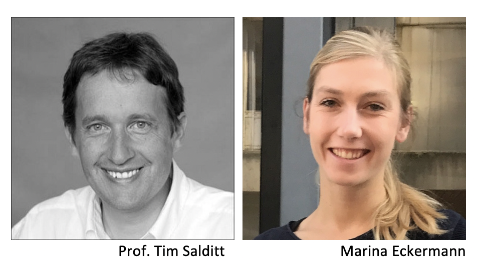
Abstract (Tim Salditt)
X‐rays can deeply penetrate matter and thus provide information about the functional (interior) architecture of complex samples, from biological tissues and cells to nanoscale composite materials. Until recently, however, this potential of hard x‐rays in view of penetration, spatial resolution, contrast, and compatibility with environmental conditions was significantly limited by the lack in suitable in x‐ray optics. With the advent of highly brilliant radiation, and the development of lens‐less diffractive imaging and coherent focusing, the situation has changed. We now have nano‐focused coherent x‐ray synchrotron beams at hand to probe nanoscale structures both in scanning and in full field imaging and tomography. We explain how the central challenge of inverting the coherent diffraction pattern can be mastered by different reconstruction algorithms in the optical far and nearfield. In particular, we present full field projection imaging at high magnification, recorded by illumination with advanced x‐ray waveguide optics [1,2], and show how imaging and diffraction can be combined to investigate biomolecular structures within biological cells [3], also correlatively with super‐resolution light microscopy. We present different examples of biophysical and biomedical applications [4,5], including 3d virtual histology of human brain tissue [5].
[1] M. Bartels, M. Krenkel, J. Habe, R.N. Wilke, T. Salditt, X‐Ray Holographic Imaging of Hydrated Biological Cells in Solution, Phys. Rev. Lett. 114, 048103 (2015).
[2] L. M. Lohse, A.‐L. Robisch, M. Töpperwien, S. Maretzke, M. Krenkel, J. Hagemann and T. Salditt A phase‐retrieval toolbox for X‐ray holography and tomography Journal of Synchrotron Radiation (2020), 27, 3
[3] M. Bernhardt, J.‐D. Nicolas, M. Osterhoff, H. Mittelstädt, M. Reuss, B. Harke, A. Wittmeier, M. Sprung, S. Köster, T. Salditt. Correlative microscopy approach for biology using x‐ray holography, x‐ray scanning diffraction, and STED microscopy Nat. Comm. (2018), 9, 3641
[4] M. Reichardt, C. Neuhaus, J‐D. Nicolas, M. Bernhardt, K. Toischer and T. Salditt X‐ray structural analysis of single adult cardiomyocytes: tomographic imaging and micro‐diffraction. Biophysical Journal (2020), 119, 7, 1309‐1323
[5] M. Töpperwien, F. Van der Meer, C. Stadelmann, and T. Salditt. Three‐dimensional virtual histology of human cerebellum by X‐ray phase‐contrast tomography, Proceedings of the National Academy of Sciences (2018), 201801678
Abstract (Marina Eckermann)
Severe progression of Covid‐19 emerges with a very diffuse, multi‐organ disease pattern, and is particularly found in lung tissue leading to lethal respiratory failure. With 3d virtual histology, we could exploit the optical properties of hard X‐rays to study the disease mechanisms on different length scales using propagation‐based phase‐contrast computed-tomography (PB‐CT). We probed post mortem autopsy lung tissue samples from Covid‐19 patients, and quantified the very diverse pathology: Thickened alveolar walls are observed, covered by hyaline membrane (see Figure). Also the vasculature is affected, showing abnormal vascular growth as sprouting or more importantly intussusceptive neoangiogenesis. Further signs of inflammation include the infiltration with lymphocytes or the formation of thrombi.
In this talk, I will present the multi‐scale PB‐CT approach based on inhouse CT and the high resolution holo‐tomography at the GINIX endstation of the P10 beamline (DESY, Hamburg), explain the data analysis workflow, and the main findings and implications for Covid‐19.
Key Reference:
M. Eckermann*, J. Frohn*, M. Reichardt*, M. Osterhoff, M. Sprung, F. Westermeier, A.Tzankov, C. Werlein, M. Kuehnel, D. Jonigk and T. Salditt “3d Virtual Patho‐Histology of Lung Tissue from Covid‐19 Patients based on Phase Contrast X‐ray Tomography”, eLife (2020); doi: 10.7554/eLife.60408
Webinar moderators
Members of the organising group.
Please contact either gerardina.carbone@maxiv.lu.se or asa.grunning@linxs.lu.se for any questions.
During our events we sometimes take photographs and short film clips to profile our activities. Please let us know if you don’t want to be in any photos/films before we start the event. Some webinars are recorded to be used for educational purposes in the LINXS website.
By registering to our events you give your permission to LINXS, according to the General Data Protection Regulation (GDPR), to register your name and e-mail address to be used for the sole purpose of distributing newsletters and communications on LINXS activities.


